




NRA and NRP: two nuclear-reaction analysis techniques, presented in detail below.
NRA (Nuclear Reaction Analysis) and NRP (Nuclear Resonance Profiling) both exploit nuclear reactions induced by a beam of light ions, protons (p) or deuterons (d), on the light nuclei of the target, to measure elements (Li, B, C, N, O, F) with an isotopic selectivity that RBS cannot offer. They are nevertheless two distinct techniques, presented separately below.
NRA, Nuclear Reaction Analysis
NRA measures light elements by identifying the particle emitted by a beam-induced nuclear reaction, rather than by analysing simple elastic scattering.
Physical principle
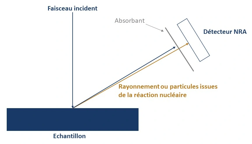
When the impact parameter of a light incident ion on a light nucleus (Z₂ < 16) is small enough, a nuclear reaction occurs: an unstable compound nucleus forms and then decays within a very short time (10⁻¹⁶ to 10⁻¹⁸ s), emitting a charged particle and/or a γ photon. The energy of the emitted particle depends on:
- The “Q” of the reaction, characteristic of the isotope-reaction pair (hence of the element and of the isotope present, unlike RBS)
- The depth at which the reaction took place (energy loss of the beam on the way in)
Technical characteristics
- Incident beam: p or d, from a few hundred keV to a few MeV
- Typical reactions: ¹⁶O(d,p)¹⁷O, ¹⁴N(d,α)¹²C
- Detector: silicon detector, with an absorber to stop backscattered incident ions and observe only the reaction products
- Depth resolution: of the order of a hundred nanometres, limited by the energy spread in the absorber
Advantages
- Isotopic selectivity, distinguishes ¹⁶O from ¹⁸O, ¹⁴N from ¹⁵N, where RBS only sees the element
- Very sensitive to light elements poorly or not detected in conventional RBS (Li, B, C, N, O, F)
- Enables isotopic tracing (e.g. ¹⁸O as an oxide growth marker)
Limitations
- Complex cross-sections, specific to each reaction: require well-characterised reference samples
- Depth resolution limited to about a hundred nm by the energy spread in the absorber
Typical applications on SAFIR
- Measurement of oxygen and carbon in thin oxide layers
- Determination of nitride stoichiometry (Si₃N₄) via reactions on ¹⁴N, ²⁸Si and ²⁹Si
- Isotopic tracing of oxide growth and oxygen diffusion
NRP, Nuclear Resonance Profiling
NRP exploits nuclear reactions exhibiting a narrow cross-section resonance to obtain depth concentration profiles with a resolution close to the nanometre, one of the best resolutions in all of nuclear microanalysis. The method was developed in the 1980s by G. Amsel and his collaborators, within the team that would give rise to SAFIR.
Physical principle
Some nuclear reactions show one or more sharp peaks (width < 2 keV, sometimes < 100 eV) in their cross-section. By scanning the incident beam energy around this resonance, emitted particles are detected only when the ions, slowed down by their path through matter, reach exactly the resonance energy at a given depth. The resulting excitation curve, the count rate as a function of beam energy, is thus a direct image of the concentration profile of the isotope of interest:
- Each beam energy probes a precise depth, given by the stopping power of the material
- The depth resolution depends on the width of the resonance and on the energy spread of the incident beam, not on the resolution of the detector, unlike RBS or conventional NRA
Technical characteristics
- Incident beam: protons or deuterons, with a very low energy spread
- Reference reaction: ¹⁸O(p,α)¹⁵N, resonance at 151 keV, full width at half maximum < 100 eV
- Depth resolution: very good depth resolution near the surface
- Analysed depth: a few hundred nanometres, with resolution degrading with depth due to the energy spread of the incident beam in matter
Advantages
- Very good depth resolution near the surface, without requiring a high-resolution detector
- Direct isotopic tracing (e.g. ¹⁸O as an oxide growth marker) with nanometre precision
- Well-established stochastic theory (Amsel et al.) allowing the concentration profile to be reconstructed from the measured excitation curve
Limitations
- Requires that a narrow, usable resonance exists for the isotope of interest; not all reactions lend themselves to it
- Requires a beam with very low energy spread to fully exploit the narrowest resonances
- Resolution degrades quickly beyond the very first nanometres/tens of nanometres
Typical applications on SAFIR
- Oxygen-18 concentration profiles in silica, via the 151 keV resonance of ¹⁸O(p,α)¹⁵N
- Fine study of the growth kinetics of ultrathin oxides, layer by layer
- Isotopic tracing of oxygen diffusion near the surface
Complementarity with other techniques
NRA and NRP complement RBS and ERDA for light elements (lithium to fluorine) that RBS distinguishes poorly, by providing an isotopic selectivity that neither of the other two techniques can offer on its own.
Reference publications
NRA, J. Siejka, J.P. Nadai, G. Amsel, “A Study of the Oxygen Growth Laws of Anodic Oxide Films on Aluminum and Tantalum Using Nuclear Microanalysis of O16 and O18”, Journal of The Electrochemical Society, vol. 118, no. 5, p. 727, 1971. DOI: 10.1149/1.2408154
NRP, A. L’Hoir, J.J. Ganem, I. Trimaille, I.C. Vickridge, “Direct measurement of molecular breakup dynamics and vicinage effect of swift proton clusters using narrow nuclear resonance profiling”, Nuclear Instruments and Methods B, 2010. See on HAL